
Maxillary Sinus Communication: Risks, Treatments, and Recovery After Extraction
How common is sinus perforation after tooth extraction? This is a frequently asked question by patients preparing for upper molar removal. In clinical terms, a sinus perforation—medically known as an oroantral communication (OAC)—occurs in approximately 1% to 5% of all maxillary (upper) posterior tooth extractions. While the overall incidence across all dental extractions is relatively low, the risk increases significantly when removing upper first and second molars due to the close anatomical proximity of their roots to the floor of the maxillary sinus. Fortunately, modern dental protocols in 2026 ensure that when these communications do occur, they are swiftly diagnosed and highly treatable.
Key Takeaways
- An oroantral communication (OAC) occurs in roughly 1% to 5% of upper posterior extractions.
- The highest risk involves the extraction of the maxillary first and second molars.
- Small perforations (under 2 millimeters) typically heal spontaneously with a normal blood clot.
- Large perforations (over 6 millimeters) usually require immediate surgical closure using specialized flap techniques.
- Strict adherence to post-operative sinus precautions (e.g., avoiding nose blowing and using decongestants) is critical for proper healing.
- Pre-operative 3D imaging, such as Cone Beam Computed Tomography (CBCT), is the 2026 standard of care for assessing risk before surgery.
Understanding the Anatomy of the Maxillary Sinus
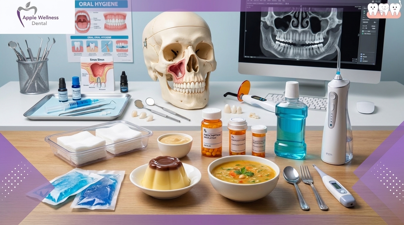
To understand why sinus perforations occur, it is essential to examine the anatomy of the upper jaw. The maxillary sinuses are large, air-filled cavities located just above the upper posterior teeth, sitting behind the cheekbones on either side of the nose. These cavities are lined by a thin respiratory membrane known as the Schneiderian membrane.
In many adult patients, the roots of the maxillary premolars and molars extend upward into the alveolar bone, separated from the sinus cavity by only a paper-thin layer of bone. In some cases, this bone is virtually absent, meaning the tooth roots are in direct contact with the sinus membrane. According to anatomical data provided by the National Institutes of Health, a phenomenon called sinus pneumatization—where the sinus cavity expands downward as people age—further increases this root-to-sinus proximity. When a tooth is extracted, this thin barrier can tear, creating an unnatural opening between the oral cavity and the sinus cavity.
Statistical Incidence: Examining the Clinical Data

While the prospect of a sinus perforation sounds alarming, clinical data provides reassuring context. Research and surgical records consistently show that OACs are a known, manageable anatomical complication rather than a routine surgical error.
Studies indicate that the incidence rate sits between 1% and 5% for upper molar extractions. The risk profile breaks down distinctly depending on the specific tooth being removed. The maxillary first molar accounts for nearly 50% of all reported sinus communications. This is followed by the maxillary second molar and the second premolar. Extractions of front teeth (incisors and canines) carry virtually zero risk of sinus perforation due to their forward position in the mouth.
Key Risk Factors for Oroantral Communication
Several anatomical and surgical factors increase the likelihood of experiencing a sinus perforation during an extraction:
- Root Morphology: Teeth with widely divergent or severely curved roots require more manipulation during extraction, increasing the stress on the surrounding bone.
- Sinus Pneumatization: As mentioned, an enlarged sinus cavity leaves less bone between the tooth apex and the sinus floor.
- Periapical Infections: Chronic infections or cysts at the root tip can destroy the intervening bone before the extraction even takes place.
- Surgical Trauma: Complex surgical extractions involving sectioning (cutting) the tooth or removing bone natively carry a higher risk of membrane disruption.
Recognizing the Signs and Symptoms
When an oroantral communication occurs, both the treating clinician and the patient may notice immediate signs. Proper identification in the dental chair allows for prompt management, drastically reducing the risk of secondary complications.
The most common immediate symptom is the passage of air or fluids between the mouth and the nose. A patient might notice that when they drink liquids, the fluid enters their nasal passage. Additionally, the dentist might observe air bubbling through the blood in the extraction socket during breathing. Other symptoms include an altered, hollow sound to the patient’s voice, bleeding from the nose on the side of the extraction, and an inability to build intraoral pressure (such as when puffing out the cheeks).
Clinical Diagnosis Methods
As standard medical guidelines published by the American Association of Oral and Maxillofacial Surgeons emphasize, “Immediate recognition and appropriate management of an oroantral communication are essential to prevent chronic sinusitis and fistula formation.” Dentists utilize several non-invasive methods to confirm a perforation.
The most common clinical test is the Valsalva maneuver. The dentist will ask the patient to pinch their nostrils shut and gently blow air through their nose. If a perforation exists, the trapped air will escape through the extraction socket, creating visible bubbles in the socket fluid. Furthermore, dentists use blunt periodontal probes to gently check the base of the socket, though this is done with extreme caution to avoid enlarging a small tear or pushing debris into the sinus cavity.
Classification and Treatment Protocols
The management of a sinus perforation is entirely dependent on its size. Dental professionals categorize OACs into three primary sizes, each requiring a specific treatment protocol to ensure predictable healing and prevent infection.
| Perforation Size | Classification | Standard Treatment Protocol |
|---|---|---|
| Under 2 mm | Small | Spontaneous healing via normal blood clot formation. Strict sinus precautions prescribed. |
| 2 mm to 6 mm | Moderate | Placement of resorbable collagen sponges or gelatin foam (Gelfoam), secured with figure-eight sutures. |
| Over 6 mm | Large | Immediate surgical closure using flap techniques (e.g., buccal advancement flap or buccal fat pad). |
Surgical Closure Techniques
For perforations larger than 6 millimeters, the body cannot reliably form a stable blood clot across the gap. In these scenarios, the clinician will perform a surgical closure to seal the socket. The Buccal Advancement Flap is the most widely utilized technique. This involves making a small incision in the gum tissue on the cheek side (buccal side) of the extraction site, stretching the tissue, and suturing it securely over the socket.
In more complex cases, or cases where a previous closure has failed, a Buccal Fat Pad graft may be utilized. This advanced technique pulls a small amount of native fat from the cheek into the surgical site. The fat pad possesses an excellent blood supply, which drastically accelerates epithelialization (tissue healing) and closes the defect with a success rate exceeding 90%.
Crucial Post-Operative Sinus Precautions
Regardless of whether the perforation was 1 millimeter or 8 millimeters, patient compliance with post-operative instructions is the single most important factor in successful healing. The overarching goal of “sinus precautions” is to prevent changes in air pressure between the nasal cavity and the mouth, which could dislodge the protective blood clot or rupture the surgical sutures.
For a minimum of 14 days following the extraction, patients must adhere to the following strict guidelines:
- Do Not Blow Your Nose: If a runny nose occurs, wipe it gently. Blowing the nose creates immense pressure that will immediately force air through the healing socket.
- Sneeze With Your Mouth Open: If sneezing is unavoidable, keeping the mouth open prevents pressure from building up in the sinus cavity.
- Avoid Straws and Smoking: The suction created by straws, cigarettes, or vaping devices can dislodge the blood clot in seconds, leading to a dry socket and a reopened perforation.
- Avoid Wind Instruments: Playing instruments like the trumpet, flute, or saxophone must be suspended during the recovery period.
- Take Prescribed Medications: Dentists typically prescribe a broad-spectrum antibiotic (like Amoxicillin or Augmentin) to prevent sinus infections, alongside systemic or topical nasal decongestants (such as pseudoephedrine) to keep the nasal passages clear and reduce membrane swelling.
Long-Term Complications of Untreated Perforations
While prompt treatment leads to excellent outcomes, an undiagnosed or ignored sinus perforation can lead to severe chronic issues. If an OAC fails to heal within 48 to 72 hours, the constant flow of oral bacteria into the sterile sinus cavity will almost inevitably cause acute sinusitis.
Over time, the cells lining the oral cavity will migrate down into the hole to meet the cells lining the sinus cavity. This creates a permanent, skin-lined tunnel known as an Oroantral Fistula (OAF). Once a fistula has formed, it will never heal on its own. It requires a more extensive surgical intervention, often accompanied by endoscopic sinus surgery performed by an Ear, Nose, and Throat (ENT) specialist to clean out chronic sinus infections. Clinical statistics indicate that nearly 50% of untreated large perforations will develop into a permanent fistula within four weeks.
Prevention Through Modern Dental Technology
In 2026, the emphasis in modern dentistry is squarely on prevention through advanced diagnostic imaging. Traditional two-dimensional X-rays, while useful, often compress complex three-dimensional anatomy into a flat image, masking the true proximity of the tooth roots to the sinus floor.
Today, the gold standard for high-risk extractions is Cone Beam Computed Tomography (CBCT). As highlighted by resources from the American Dental Association, CBCT scans provide a hyper-accurate, 3D roadmap of the patient’s exact anatomy. If a scan reveals roots protruding into the sinus cavity, the dental surgeon can pre-plan a surgical extraction. By meticulously sectioning the tooth into smaller pieces and removing them individually, the surgeon drastically reduces the rotational forces applied to the surrounding bone, significantly mitigating the risk of tearing the Schneiderian membrane.
Conclusion
A sinus perforation after a tooth extraction is a known anatomical risk, primarily associated with the removal of upper molars. With an occurrence rate of roughly 1% to 5% in maxillary posterior extractions, it is a relatively common complication that oral health professionals are well-equipped to handle. Through immediate clinical diagnosis, appropriate surgical or conservative management, and strict adherence to post-operative sinus precautions, patients can expect a full, uneventful recovery without long-term sinus issues.
If you are experiencing symptoms of a sinus communication—such as air escaping through a healing socket or liquids passing into your nose—immediate evaluation is crucial. To ensure your oral health is protected and to receive an expert assessment, contact our dental office today to schedule a comprehensive examination.
References
- American Association of Oral and Maxillofacial Surgeons (AAOMS). Clinical Guidelines on Management of Oroantral Communications.
- American Dental Association (ADA). Guidelines on the Use of Cone Beam Computed Tomography in Exodontia.
- National Institutes of Health (NIH). Research on Maxillary Sinus Pneumatization and Root Morphology.
- Mayo Clinic. Clinical Information on Sinusitis and Oroantral Fistulas.
- Hupp, J.R. Contemporary Oral and Maxillofacial Surgery. (Textbook data regarding anatomical risks and surgical flap techniques).