
Comprehensive Guide on Managing Neuropathy Following Dental Extraction
How to treat nerve pain after tooth extraction is a clinical challenge that requires immediate evaluation, a combination of pharmacological therapies, and sometimes microsurgical intervention. While dental extractions—particularly the removal of mandibular third molars (wisdom teeth)—are routine procedures in 2026, complications involving the complex network of facial nerves occasionally occur. When the inferior alveolar nerve or lingual nerve sustains trauma during surgery, patients may experience an array of uncomfortable sensations, ranging from numbness to severe, burning neuropathic pain. Addressing this neuropathy swiftly is critical, as early intervention significantly improves the likelihood of complete neural regeneration and sensory recovery.
Key Takeaways
- Immediate Action: Medical evaluation within the first 48 hours is crucial for minimizing permanent nerve damage.
- First-Line Treatments: High-dose corticosteroids and non-steroidal anti-inflammatory drugs (NSAIDs) are the primary pharmacological defenses against acute nerve inflammation.
- Surgical Window: If severe symptoms persist, microsurgical repair is most effective when performed between 3 and 6 months post-injury.
- High Recovery Rates: Approximately 85% of post-extraction nerve injuries resolve spontaneously within 8 weeks.
- Nutritional Support: Vitamin B complex (specifically B1, B6, and B12) is scientifically proven to aid in peripheral nerve regeneration.
Understanding Trigeminal Nerve Injuries in Dentistry

The human jaw is heavily innervated by branches of the trigeminal nerve. During lower tooth extractions, two specific branches are at risk: the inferior alveolar nerve (IAN), which runs through the jawbone directly beneath the tooth roots, and the lingual nerve (LN), which supplies sensation to the tongue. Research from the National Institute of Dental and Craniofacial Research indicates that the proximity of these nerves to the roots of wisdom teeth makes them susceptible to mechanical trauma.
Dental professionals classify nerve injuries using Seddon’s classification system. Neuropraxia is a mild stretch or bruise to the nerve where the outer sheath remains intact, typically resulting in temporary numbness. Axonotmesis involves damage to the internal nerve fibers but an intact outer structure, requiring a longer recovery period. The most severe classification, Neurotmesis, constitutes a complete severing of the nerve, which invariably requires surgical intervention for any hope of sensory return. Clinical data demonstrates that temporary nerve injury occurs in roughly 1% to 5% of all lower wisdom tooth extractions, whereas permanent injury lasting more than six months occurs in less than 1% of cases.
Recognizing the Symptoms of Nerve Damage
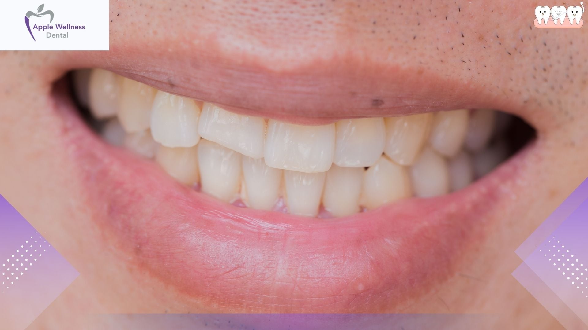
Identifying the exact nature of sensory dysfunction is the first step in formulating a treatment plan. Nerve damage does not always manifest strictly as pain; it presents through a spectrum of altered sensations known as neuropathy. Patients must track their symptoms closely and report them accurately to their oral surgeon.
- Paresthesia: This is a painless, altered sensation often described as numbness, tingling, or a “pins and needles” feeling, similar to the sensation of local anesthetic wearing off.
- Dysesthesia: This represents an unpleasant and often painful abnormal sensation. Patients describe it as burning, shocking, or an intense itching deep within the tissue.
- Hyperalgesia: An exaggerated pain response to a mildly painful stimulus. A simple tap on the chin or a mild temperature change can trigger severe discomfort.
- Allodynia: Experiencing pain from stimuli that normally do not provoke pain, such as the light touch of a toothbrush or wind blowing against the face.
Immediate Steps: What to Do in the First 48 Hours
Time is of the essence when treating iatrogenic nerve injuries. The inflammatory response that follows surgical trauma can compress the nerve within the narrow bony canal of the jaw, exacerbating the initial injury. Taking immediate, structured action minimizes secondary damage.
- Contact the Operating Surgeon Immediately: Do not wait for a scheduled follow-up appointment. Inform the clinic that the local anesthesia has worn off but numbness or shooting pain persists.
- Initiate Anti-Inflammatory Therapy: Upon a doctor’s recommendation, beginning a regimen of NSAIDs (such as ibuprofen) helps reduce local tissue swelling. In highly suspected cases of direct trauma, a dentist will prescribe a short, tapering dose of oral corticosteroids (like Dexamethasone).
- Apply Cold Compresses: In the first 48 hours, applying ice packs to the exterior of the jaw for 20-minute intervals restricts blood flow, effectively reducing the edematous swelling that compresses the inferior alveolar nerve.
- Avoid Aggressive Chewing: Eat a strict liquid or ultra-soft diet to prevent accidental biting of the numb lip, cheek, or tongue, which can cause severe lacerations.
Pharmacological Treatments and Medications
When physical symptoms transition from acute postoperative swelling to chronic neuropathic pain, the treatment protocol shifts toward specialized pain management. Standard over-the-counter analgesics like acetaminophen are largely ineffective against neuropathic pain, as the pain originates from nerve misfiring rather than standard tissue inflammation. Consequently, oral and maxillofacial surgeons utilize specific classes of prescription medications to stabilize nerve membranes.
| Medication Class | Common Examples | Mechanism of Action | Expected Outcome |
|---|---|---|---|
| Corticosteroids | Dexamethasone, Prednisone | Reduces acute neural inflammation and prevents edema within the nerve canal. | Prevents secondary compression damage during the first week post-op. |
| Anticonvulsants | Gabapentin, Pregabalin | Decreases the release of excitatory neurotransmitters in the central nervous system. | Provides a 30% to 50% reduction in burning, shooting dysesthesia. |
| Tricyclic Antidepressants | Amitriptyline, Nortriptyline | Alters pain processing pathways in the brain by increasing serotonin and norepinephrine. | Improves sleep and dampens chronic neuropathic throbbing. |
| Topical Anesthetics | Lidocaine patches, Capsaicin cream | Desensitizes localized nerve endings in the affected soft tissue. | Offers temporary, localized relief for allodynia on the lip or chin. |
According to the World Health Organization guidelines on pain management, tailoring the specific medication to the patient’s exact symptom profile ensures the highest efficacy while minimizing side effects. Gabapentinoids, for instance, are currently considered the gold standard for managing dysesthesia following dental surgery.
Advanced Therapies and Surgical Interventions
If pharmacological interventions yield insufficient results and sensory deficits show no signs of improvement after three months, advanced clinical therapies are deployed. One non-invasive modality gaining widespread traction in 2026 is Low-Level Laser Therapy (LLLT), also known as photobiomodulation. By applying targeted infrared light to the injured tissue, LLLT stimulates cellular metabolism, increases local blood flow, and accelerates the regeneration of Schwann cells, which are responsible for nerve fiber myelination.
When imaging (such as CBCT or MRI) confirms physical severing or severe entrapment of the nerve, microsurgery is required. Surgical options include nerve decompression (removing bone or scar tissue pressing on the nerve), neurorrhaphy (direct suturing of severed nerve ends), and the placement of nerve allografts. The timing of this surgery is a critical determinant of success.
As Dr. Tara Renton, a globally recognized researcher in trigeminal nerve injuries, emphasizes in her published research: “Early intervention and accurate mapping of the neuropathy are essential for maximizing the potential for neural recovery.”
Furthermore, timing dictates the biological capacity for regeneration. According to Dr. John Zuniga, a leading Professor of Oral and Maxillofacial Surgery: “The window for optimal surgical repair of the trigeminal nerve is typically between three and six months post-injury, after which the potential for meaningful sensory recovery begins to decline dramatically due to Wallerian degeneration.”
Dr. Vincent Ziccardi, Chief of Oral and Maxillofacial Surgery at Rutgers School of Dental Medicine, corroborates this standard of care, stating: “Microsurgical intervention remains a highly predictable treatment for severe nerve injuries when non-surgical therapies fail to produce sensory improvement within the first three months.”
Holistic and Supportive Management
Beyond traditional Western medicine, comprehensive nerve recovery protocols incorporate targeted nutritional supplementation. Peripheral nerves possess a unique capability to regenerate, growing at an average rate of 1 to 2 millimeters per day. This complex biological process requires specific metabolic building blocks.
Clinical nutritionists strongly advocate for high doses of the Vitamin B complex. Specifically, Vitamin B1 (Thiamine) provides the cellular energy required for nerve repair, Vitamin B6 (Pyridoxine) facilitates neurotransmitter synthesis, and Vitamin B12 (Cobalamin) is strictly essential for remyelination—the rebuilding of the nerve’s protective outer sheath. Deficiencies in any of these vitamins fundamentally stall the healing process.
Additionally, chronic facial pain frequently takes a severe psychological toll on patients, leading to anxiety and depression. Integrating Cognitive Behavioral Therapy (CBT) into the recovery plan equips patients with pain-coping mechanisms, ensuring that the psychological burden of neuropathy is actively managed alongside the physical symptoms. Acupuncture has also demonstrated moderate efficacy in stimulating nerve pathways and providing natural analgesic effects through the release of endorphins.
Recovery Timeline and Prognosis
Patience is heavily required when dealing with nerve injuries. The physiological reality is that nerves heal much slower than muscle or bone. Statistical data from the American Association of Oral and Maxillofacial Surgeons reveals that approximately 85% of inferior alveolar nerve injuries resolve spontaneously within 8 weeks. For the remaining 15%, the timeline extends into months. If a patient experiences “formication”—a tingling sensation that feels like ants crawling just beneath the skin—this is typically celebrated by surgeons as a positive clinical indicator of active neural regeneration. Conversely, if zero sensory improvement is noted by the 12-week mark, permanent deficits become highly probable without surgical intervention.
Frequently Asked Questions
How long does nerve pain last after a tooth extraction?
For the vast majority of patients, nerve pain and temporary numbness resolve within 4 to 8 weeks. However, if the nerve sustained severe trauma or was completely severed, pain and sensory deficits can become permanent without specialized surgical intervention.
Can an X-ray show if my nerve is damaged?
Standard 2D dental X-rays cannot show soft tissue like nerves. However, a 3D Cone Beam Computed Tomography (CBCT) scan or a specialized MRI can reveal if bone fragments, root tips, or surgical hardware are compressing the nerve canal.
Does Vitamin B12 actually help repair dental nerves?
Yes, Vitamin B12 (specifically in the form of methylcobalamin) is scientifically proven to assist in the remyelination of damaged peripheral nerves. It provides the biological materials necessary to rebuild the protective sheath around the nerve fibers.
When is it too late for nerve repair surgery?
The optimal window for trigeminal nerve microsurgery is between 3 and 6 months post-injury. Surgeries performed after 12 months have a significantly lower success rate due to permanent scar tissue formation and irreversible nerve degeneration.
Are Gabapentin side effects worth the pain relief?
For patients suffering from severe, sleep-disrupting dysesthesia, Gabapentin is highly effective. While it can cause temporary dizziness and fatigue, physicians monitor dosages closely to balance pain relief with manageable side effects.
Conclusion
Experiencing neuropathy after dental surgery is a stressful and painful ordeal, but advancements in diagnostic imaging, targeted pharmacology, and microsurgical techniques provide robust solutions in 2026. By understanding the anatomy involved, recognizing symptoms early, and adhering to the 3-to-6-month surgical evaluation window, patients maximize their chances for complete sensory recovery. If you are experiencing persistent burning, tingling, or numbness following an extraction, do not adopt a wait-and-see approach. Seek immediate professional evaluation to map your injury and initiate a tailored recovery plan. Contact us today to schedule a comprehensive diagnostic consultation with our specialists.
References
- American Dental Association (ADA) – Guidelines on postoperative complications and pain management.
- National Institute of Dental and Craniofacial Research – Research data on trigeminal nerve trauma and regeneration.
- American Association of Oral and Maxillofacial Surgeons (AAOMS) – Clinical statistics on third molar extraction outcomes.
- World Health Organization – Global guidelines on the pharmacological management of neuropathic pain.