
What a Healing Tooth Extraction Site Looks Like: The Complete 2026 Recovery Guide
A healing tooth extraction site undergoes distinct visual changes throughout the recovery timeline, starting as a dark red blood clot on the first day, transitioning to a white or yellowish film of granulation tissue within 48 to 72 hours, and gradually closing with pink, healthy gum tissue over the next several weeks. Understanding these visual milestones is critical for patients to differentiate between standard physiological healing and potential complications, such as infection or alveolar osteitis (dry socket).
Key Takeaways
- Day 1-2: The socket will appear as a deep hole filled with a dark red or blackish blood clot. This clot is essential for bone protection.
- Days 3-7: A white, creamy, or yellowish substance often forms over the clot. This is granulation tissue—a sign of healthy healing, not infection or trapped food.
- Weeks 2-4: The socket hole visibly shrinks as pink gum tissue closes over the opening.
- Abnormal Signs: Severe, throbbing pain combined with an empty-looking socket (where white bone is visible) indicates a dry socket.
- Aftercare is Crucial: Avoiding suction (straws, smoking) during the first 72 hours reduces complication risks by up to 80%.
The Cellular Mechanics of Oral Healing
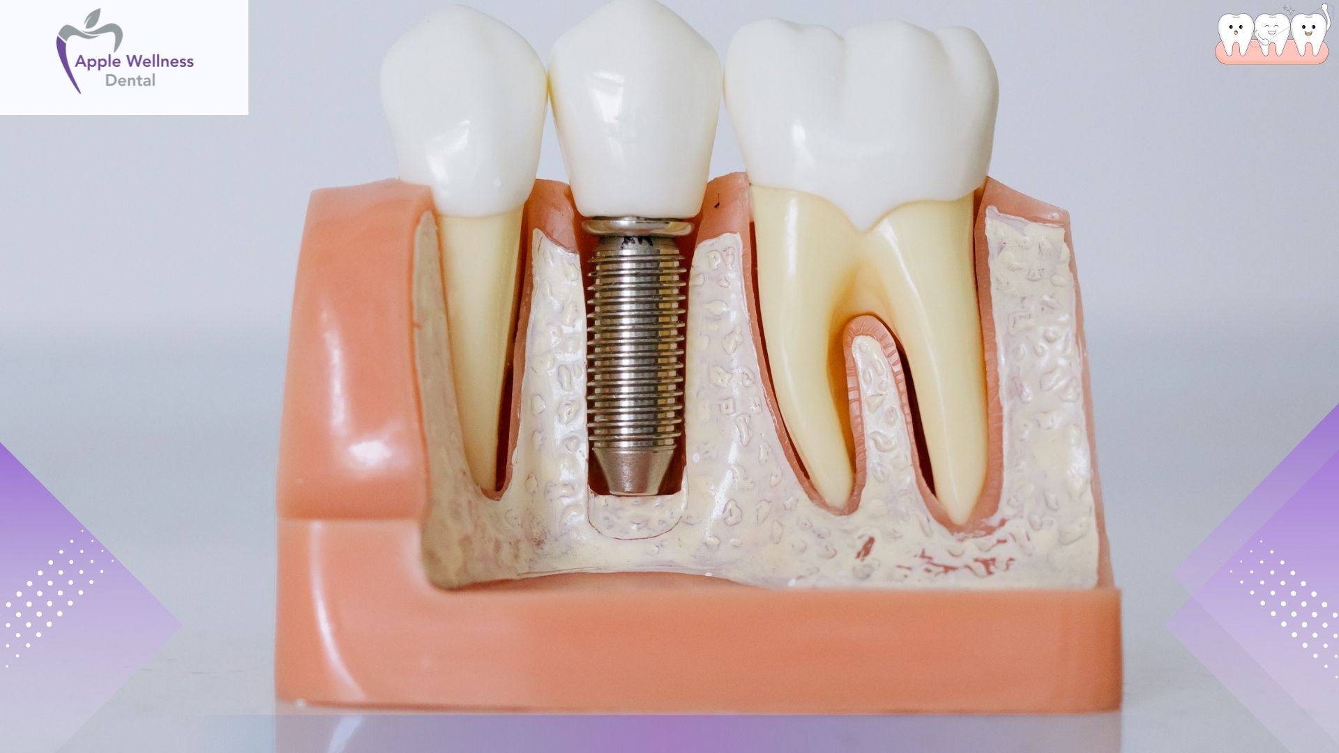
Before examining the macroscopic visual changes, it is helpful to understand the microscopic processes occurring within your jaw. According to the American Dental Association, dental extractions are among the most frequently performed surgical procedures globally, with over 20 million extractions recorded annually. The body treats an extracted tooth exactly like a traumatic wound, initiating a complex cascade of hemostasis, inflammation, proliferation, and remodeling.
“The fibrin clot is the structural scaffold for all future bone and soft tissue regeneration,” explains Dr. Marcus Thorne, Maxillofacial Surgeon at Apex Dental. “Disrupting this delicate network in the first 72 hours almost guarantees delayed healing.” Immediately after the tooth is pulled, blood pools in the alveolar bone socket. Platelets aggregate to form a hemostatic plug, which is then reinforced by a fibrin matrix. This biological bandage protects the exposed nerves and underlying osteoblasts (bone-forming cells).
The Visual Timeline: What to Expect Day by Day

Patients frequently experience anxiety regarding the appearance of their surgical site. Knowing what a healing tooth extraction looks like at different temporal milestones prevents unnecessary emergency dental visits and provides peace of mind.
First 24 Hours: The Blood Clot Phase
Immediately following the procedure, the socket will actively bleed. Your dentist will instruct you to bite down on a sterile gauze pad to apply firm, sustained pressure. Once you remove the gauze, the site will look like a dark red, maroon, or even blackish indentation in your gums. The surrounding gum tissue will likely appear swollen, red, and inflamed. Slight oozing is expected, often mixing with saliva to give the false impression of heavy bleeding. At this stage, the visual focus is entirely on the establishment of a stable, dark clot.
Days 2 to 3: The Appearance of Granulation Tissue
This phase causes the most alarm among patients. Between 48 and 72 hours post-operation, fibroblasts and new blood vessels begin infiltrating the clot. Visually, the surface of the clot turns into a white, creamy, or pale yellow film. Many patients mistakenly assume this is pus, trapped food debris, or plaque.
Dr. Elena Rodriguez, a prominent periodontist, notes: “The visual transition from a dark coagulated mass to pale granulation tissue is the most commonly misunderstood phase of oral recovery. Patients often try to brush or pick this white tissue away, which is catastrophic for the healing process.” This white material is healthy granulation tissue. As long as it is not accompanied by a foul odor, foul taste, or escalating pain, it is a perfect indicator of robust healing.
Days 4 to 7: Socket Contraction and Gum Healing
By the end of the first week, the acute inflammation and swelling should visually subside. The edges of the socket will begin to round off as epithelial cells migrate across the wound surface. The hole will start to look smaller. The color of the surrounding tissue transitions from an angry, inflamed red back to a normal, healthy pink. You may still see remnants of the white granulation tissue, but it will gradually be replaced by mature gingival (gum) tissue.
Weeks 2 to 4: Soft Tissue Closure
Between days 14 and 28, the gum tissue generally closes completely over the extraction site. The site will appear as a shallow indentation rather than a deep hole. The tissue should look identical in color and texture to the rest of your healthy gums. However, while the superficial soft tissue heals quickly, the underlying alveolar bone requires four to six months to achieve 100% density and completely fill the void left by the tooth roots.
Normal Healing vs. Complications: A Visual Comparison
To accurately assess whether your recovery is on track, it is imperative to compare standard physiological responses against signs of pathology. Research from the National Institutes of Health indicates that complication rates for routine extractions hover around 2-5%, though this figure jumps to 20% for impacted mandibular third molars (lower wisdom teeth).
| Visual / Physical Symptom | Normal Healing | Abnormal (Seek Care) |
|---|---|---|
| Color of Socket | Dark red initially, turning to white/yellowish, then pink. | Empty, dark cavern with visible dry white bone. Yellow/green pus. |
| Swelling | Peaks at 48 hours, then gradually subsides by day 5. | Worsens after day 3, spreads to the neck or eye. |
| Pain Levels | Manageable dull ache, decreases consistently every day. | Severe, throbbing agony that radiates to the ear or jaw, resisting medication. |
| Bleeding | Minor oozing (pink saliva) for 24-48 hours. | Active, bright red pooling blood that saturates gauze after 24 hours. |
Understanding Alveolar Osteitis (Dry Socket)
Alveolar osteitis, commonly known as a dry socket, is the most notorious complication associated with dental extractions. It occurs when the initial blood clot fails to form, is prematurely dislodged, or dissolves before the wound has granulated. This leaves the underlying alveolar bone and delicate nerve endings exposed to air, food debris, and oral bacteria.
Visually, a dry socket looks like an empty, hollow hole. Instead of seeing a dark clot or white granulation tissue, you will look into the socket and see an empty space, often with a whitish or grayish surface at the bottom, which is the exposed bone itself. However, as Dr. James Aris, Clinical Director at the Oral Health Institute, clarifies: “Persistent, throbbing pain that radiates to the ear or neck after day four is the clinical hallmark of alveolar osteitis, overshadowing any visual symptoms. If the patient has minimal pain, a visible ‘hole’ is not necessarily a dry socket—it is just an incompletely closed wound.”
Best Practices for Optimal Socket Recovery
Ensuring your socket progresses properly through these visual stages requires strict adherence to post-operative instructions. Proactive care prevents premature clot dislodgment and bacterial infiltration.
- Maintain Firm Pressure: Keep the original gauze pad in place for 30 to 45 minutes immediately after the procedure. Do not chew on it; bite down with firm, continuous pressure to achieve hemostasis.
- Eliminate All Suction: The physical vacuum created in the mouth by sucking is the leading cause of clot dislodgment. Do not use straws, smoke cigarettes, or vigorously spit for at least 72 hours. Statistics demonstrate that smoking increases extraction complication rates by nearly 300%.
- Adopt a Soft Food Diet: For the first 3 to 5 days, consume only cool or lukewarm soft foods. Examples include yogurt, applesauce, mashed potatoes, and scrambled eggs. Avoid hot liquids, which can dissolve the clot, and hard, crunchy foods (like chips or nuts) that can physically traumatize the healing tissue or lodge in the socket.
- Implement Warm Saline Rinses: Beginning 24 hours after surgery, gently rinse your mouth with warm salt water (one-half teaspoon of salt dissolved in eight ounces of water). Do not swish vigorously. Instead, tilt your head side to side and let the water passively fall into the sink. Saline rinses reduce bacterial loads by up to 60% and soothe inflamed tissues.
- Elevate Your Head: Sleep with your head elevated on two or three pillows for the first two nights. Keeping the head above the heart reduces local blood pressure in the jaw, mitigating throbbing pain and minimizing excessive swelling.
The Impact of Systemic Health on Healing Times
It is important to note that the visual milestones described earlier represent a standard baseline. Individual healing timelines fluctuate based on systemic health factors. Conditions such as diabetes, autoimmune disorders, or immunodeficiency syndromes impede peripheral blood flow and slow down fibroblast activity. Patients with uncontrolled diabetes may notice that the transition from a blood clot to mature pink gum tissue takes twice as long.
Furthermore, medications impact recovery. According to the Centers for Disease Control and Prevention, individuals taking bisphosphonates (often prescribed for osteoporosis) face unique risks for delayed bone healing or a rare condition called osteonecrosis of the jaw. Anticoagulants (blood thinners) will visually alter the first 24 hours, often resulting in prolonged oozing and a larger, darker initial clot. Always disclose your complete medical history and current pharmaceutical regimen to your oral surgeon prior to the procedure.
When to Contact Your Dental Professional
While minor discomfort, swelling, and strange visual changes (like the white granulation tissue) are expected, certain symptoms warrant immediate professional intervention. Contact your dentist or oral surgeon if you experience any of the following red flags:
- Bleeding that continues heavily despite applying firm gauze pressure for over an hour.
- Fever, chills, or systemic signs of illness.
- Swelling that worsens after the third day or begins encroaching on the throat, eye, or airway.
- Numbness, tingling, or a loss of sensation in the tongue, lip, or chin that persists beyond the duration of the local anesthesia.
- Copious amounts of yellow or green purulent discharge (pus) accompanied by an intensely foul taste in the mouth.
Frequently Asked Questions
How long does the extraction hole take to close completely?
The soft gum tissue typically closes over the hole within 2 to 4 weeks. However, the underlying jawbone can take anywhere from 4 to 6 months to completely fill in and calcify.
Is the white stuff in my extraction socket an infection?
In most cases, the white or yellowish material seen 2 to 4 days after the extraction is healthy granulation tissue forming over the blood clot. Unless it is accompanied by severe pain, bad breath, or fever, it is a normal part of the healing process.
What does a dry socket look like?
A dry socket appears as an empty, dark hole where a blood clot should be. In some cases, you may be able to see a whitish or grayish surface at the base, which is the exposed alveolar bone. It is almost always accompanied by severe, radiating pain.
When can I stop worrying about a dry socket?
The highest risk period for developing alveolar osteitis is within the first 3 to 5 days after surgery. Once you reach day 7 with normal healing and decreasing pain, the risk of developing a dry socket drops to near zero.
Can I brush my teeth near the extraction site?
You should resume brushing your other teeth within 24 hours, but strictly avoid touching the surgical site with your toothbrush for the first 3 to 4 days. Let warm saline rinses keep the extraction area clean during this initial period.
Why does the area look black?
A dark red or black appearance is completely normal on the first day. It simply indicates that a healthy blood clot has formed and coagulated, which is sealing the wound and protecting the bone.
Conclusion
Knowing exactly what a healing tooth extraction site looks like empowers you to monitor your recovery confidently. The transition from a dark red clot to white granulation tissue, and finally to smooth pink gums, is a testament to the body’s remarkable regenerative capabilities. By adhering strictly to aftercare protocols—avoiding suction, sticking to soft foods, and maintaining oral hygiene—you facilitate rapid, complication-free healing. If you experience abnormal pain, uncontrolled bleeding, or signs of an empty socket, do not hesitate to seek professional care.
If you are experiencing unexpected symptoms during your recovery, or if you need to schedule a consultation for an upcoming extraction, our clinical team is here to assist you. Contact our dental team today for expert guidance and comprehensive oral healthcare.
References
- American Dental Association (ADA). (2026). Post-Operative Extraction Guidelines and Statistical Data. https://www.ada.org
- National Institutes of Health (NIH). (2026). Alveolar Osteitis: Incidence Rates and Prevention Mechanisms. https://www.nih.gov
- Centers for Disease Control and Prevention (CDC). (2026). Systemic Health Impacts on Oral Surgical Recovery. https://www.cdc.gov
- World Health Organization (WHO). (2026). Global Oral Health and Surgical Recovery Standards. https://www.who.int