
Navigating Maxillary Posterior Extractions: Understanding Sinus Cavity Root Displacement
Tooth extraction when root is in sinus cavity is a highly specialized clinical scenario that requires precise surgical management. This situation, often referred to as oroantral communication (OAC) or root displacement, occurs when the bony barrier between the upper jaw (maxilla) and the maxillary sinus is breached during the removal of a tooth. When an upper molar or premolar root sits exceptionally close to, or actually protrudes into, the sinus floor, standard extraction forces can cause a pathway to open between the oral cavity and the sinus. In more complex cases, a fractured root fragment may be pushed upward into the sinus membrane. The definitive medical response involves immediate radiographic assessment—typically using 3D Cone Beam Computed Tomography (CBCT)—followed by specialized surgical retrieval techniques and the application of soft tissue grafts to close the defect. Immediate closure and strict adherence to post-operative “sinus precautions” are critical to preventing long-term complications such as chronic sinusitis or an oroantral fistula.
Key Takeaways
- High-Risk Anatomy: The maxillary first and second molars are the teeth most frequently involved in sinus complications due to their anatomical proximity to the sinus floor.
- Incidence Rates: Oroantral communications occur in approximately 0.3% to 4.7% of all maxillary posterior extractions.
- Size Dictates Treatment: Openings smaller than 2mm may heal naturally with a blood clot, while defects larger than 3mm require surgical flaps.
- Modern Diagnostics: In 2026, standard protocol dictates using CBCT imaging to precisely locate any displaced root fragment before surgical retrieval.
- Strict Post-Op Care: Patients must strictly avoid nose-blowing, using straws, or smoking for at least two weeks to protect the delicate surgical closure.
Understanding the Anatomy: The Maxillary Sinus and Upper Teeth
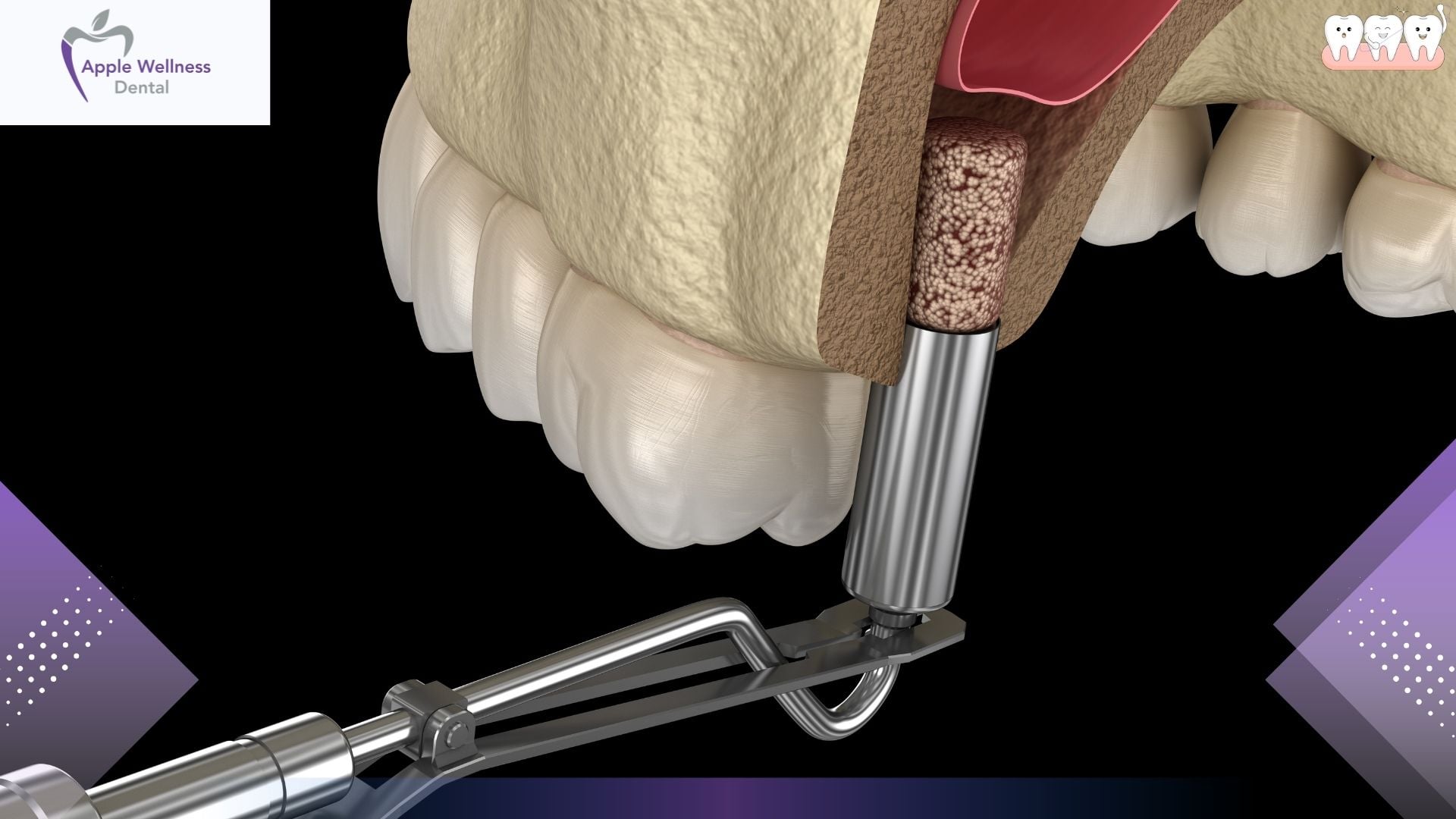
To understand why a dental root can end up in the sinus cavity, one must first examine the intricate anatomy of the human skull. The maxillary sinuses are air-filled spaces located just behind the cheekbones, directly above the upper teeth. These cavities serve multiple functions, including lightening the weight of the skull and humidifying the air we breathe. They are lined with a delicate mucosal layer known as the Schneiderian membrane.
In many individuals, the roots of the upper posterior teeth—specifically the premolars and molars—extend deeply into the alveolar bone of the maxilla. In some cases, the apices (tips) of these roots are separated from the sinus cavity by only a paper-thin layer of bone, or no bone at all, leaving the roots in direct contact with the Schneiderian membrane. Research supported by the National Institutes of Health indicates that the palatal root of the maxillary first molar is anatomically closest to the sinus floor, making it the most likely candidate for accidental sinus involvement during exodontia (tooth removal).
Why Do Tooth Roots Enter the Sinus Cavity?

During a routine extraction, a dentist applies controlled force to luxate (loosen) the tooth. However, several factors can increase the risk of breaching the sinus or displacing a root:
- Root Morphology: Divergent, curved, or exceptionally long roots can anchor tightly into the bone, requiring more force for extraction.
- Bone Resorption and Age: As patients age, the maxillary sinus can pneumatize (expand), which thins the bone separating the sinus from the tooth roots.
- Periapical Pathology: Chronic dental infections, such as abscesses at the tip of a root, can destroy the intervening bone before the extraction even begins.
- Root Fractures: If a tooth is heavily decayed or brittle, the root tip may snap off during the procedure. Attempting to retrieve a tiny 2mm fragment with standard dental elevators can inadvertently push the fragment upward through the thin sinus floor.
Signs of Oroantral Communication During Extraction
Oral and maxillofacial surgeons are trained to immediately recognize the signs of an oroantral communication (OAC) or root displacement. Prompt diagnosis in the surgical chair dictates the entire course of treatment. Common clinical signs include:
- The Valsalva Maneuver Test: The surgeon will ask the patient to pinch their nose closed and gently attempt to blow air through it. If there is a hole connecting the sinus to the mouth, the surgeon will see air bubbles or blood bubbling from the empty tooth socket.
- Voice Resonance Changes: A large defect may temporarily alter the resonance of the patient’s voice, giving it a nasal quality.
- Fluid Passage: The patient may report that fluids they drink are coming out of their nose.
- Radiographic Evidence: If a root fractures and is no longer visible in the socket, an immediate radiograph will often show the radiopaque root fragment floating above the socket within the sinus space.
The Clinical Procedure: Handling Sinus Exposures
When an oroantral communication occurs, the clinician must pivot from a standard extraction to a specialized surgical protocol. The standard of care involves the following meticulously executed steps:
- Radiographic Localization: The priority is establishing exactly where the root fragment is located. Advanced 3D imaging (CBCT) provides a millimeter-accurate map of the root’s location, ensuring no blind surgical exploration occurs.
- Retrieval Strategy: If the fragment is trapped under the sinus membrane but has not torn it, it may be retrieved carefully through the widened tooth socket using specialized micro-instruments or suction.
- The Caldwell-Luc Approach: If the root has fully breached the membrane and is lost deep within the sinus cavity, a Caldwell-Luc procedure may be necessary. This involves making a small window in the anterior maxilla (above the premolars) to access the sinus directly, locate the fragment, and flush it out.
- Endoscopic Retrieval: In modern 2026 surgical environments, functional endoscopic sinus surgery (FESS) principles are often employed. A tiny camera is inserted to locate and extract the root with minimal tissue disruption.
Surgical Defect Closure Protocols
Once the tooth and any displaced fragments are removed, the resulting hole must be managed. The clinical response is entirely dependent on the diameter of the breach. The American Dental Association emphasizes that unmanaged sinus communications frequently lead to chronic sinus infections. The table below outlines standard closure protocols based on defect size.
| Defect Size | Clinical Treatment Protocol | Prognosis |
|---|---|---|
| Under 2 mm | Encourage natural blood clot formation; place resorbable gelatin sponge (Gelfoam); suture over socket. | Excellent. Usually heals spontaneously with standard sinus precautions. |
| 2 mm to 6 mm | Requires structural support. Use of figure-eight sutures over a collagen plug or synthetic bone graft to stabilize the clot. | Highly favorable if the patient strictly adheres to post-operative instructions. |
| 7 mm or Larger | Mandates a surgical soft tissue flap (Buccal Advancement Flap, Palatal Rotational Flap, or Buccal Fat Pad graft) for primary closure. | Requires an extended healing period. High success rate when performed by a skilled surgeon. |
Advanced Grafting: The Buccal Fat Pad
For extensive sinus floor defects, one of the most reliable surgical techniques is the mobilization of the buccal fat pad (BFP). Situated in the cheek, this encapsulated mass of adipose tissue has an excellent, rich blood supply. A surgeon can gently tease this fat pad out and stretch it over the socket opening, suturing it to the palatal gums. Over the course of 3 to 4 weeks, this fat pad epithelizes—meaning it transforms into normal, healthy gum tissue—creating a permanent, waterproof seal between the mouth and the sinus.
Post-Operative Care and Sinus Precautions
Surgical intervention is only half of the solution. The patient’s adherence to post-operative instructions is the determining factor in successful healing. When a sinus breach occurs, the patient will be placed on strict “Sinus Precautions” for a minimum of 10 to 14 days.
- Do Not Blow Your Nose: Blowing the nose creates intense positive pressure within the sinus cavity, which can literally blow the blood clot or surgical graft out of the healing socket. If the patient must sneeze, they are instructed to do so with their mouth wide open to dissipate the pressure.
- Avoid Straws and Spitting: Sucking on a straw creates negative pressure in the oral cavity, which can pull the delicate clot down out of the socket.
- Avoid Smoking: Combustible tobacco products introduce extreme heat, toxic chemicals, and pressure changes that drastically reduce blood flow and increase the risk of graft failure and infection.
- No Heavy Lifting: Strenuous exercise or bearing down increases intracranial and sinus pressure, jeopardizing the surgical site.
In addition to behavioral restrictions, pharmaceutical support is standard. According to guidelines from the American Association of Oral and Maxillofacial Surgeons, clinicians routinely prescribe a broad-spectrum antibiotic (like Amoxicillin-Clavulanate) to prevent sinus infections. Furthermore, systemic decongestants (such as Pseudoephedrine) and topical nasal sprays (like Oxymetazoline) are prescribed to keep the sinus ostium open, allowing fluid to drain naturally into the nasal cavity rather than building up pressure over the extraction site.
Potential Complications: Oroantral Fistula (OAF) and Sinusitis
Failure to properly diagnose an OAC, or a patient’s failure to follow sinus precautions, can lead to chronic complications. If the opening between the mouth and the sinus does not close, the epithelial tissue from the gums will migrate down and merge with the sinus membrane lining, creating a permanent, skin-lined tunnel known as an Oroantral Fistula (OAF).
“The development of an oroantral fistula introduces a permanent highway for oral bacteria to enter the sterile environment of the maxillary sinus, inevitably leading to chronic, refractory sinusitis until surgical excision of the fistulous tract is achieved.”
Symptoms of an established OAF include a foul taste in the mouth (from sinus drainage), chronic facial pain, localized swelling, and the regurgitation of liquids through the nose. Correcting an OAF is significantly more difficult than closing an acute OAC, as the surgeon must completely excise the diseased, epithelized tract before a new flap can be attempted.
Frequently Asked Questions
Can a tooth root in the sinus cause a brain infection?
While extremely rare, untreated chronic infections in the maxillary sinus can spread to adjacent structures. However, modern antibiotic protocols and immediate surgical retrieval make this risk nearly nonexistent in contemporary dental practice.
How long does it take for a sinus communication to heal?
Small openings under 2mm typically heal within 2 to 3 weeks with proper clot stability. Larger defects repaired with surgical flaps may take 4 to 6 weeks for complete tissue integration and bone regeneration.
Will I be asleep for a displaced root retrieval?
Most simple root retrievals can be done under local anesthesia in the dental chair. However, for deep displacements requiring a Caldwell-Luc approach or endoscopic surgery, intravenous (IV) sedation or general anesthesia is usually recommended for patient comfort.
Can my general dentist fix this, or do I need a specialist?
Many experienced general dentists can manage minor sinus exposures (under 2mm) using collagen plugs and sutures. However, retrieving a deeply displaced root or performing complex flap closures typically requires referral to an Oral and Maxillofacial Surgeon.
What should I do if water comes out of my nose after an extraction?
This is a hallmark sign of an oroantral communication. Stop drinking fluids that might enter the sinus, avoid blowing your nose, and contact your treating dentist or surgeon immediately for a follow-up evaluation.
Conclusion
The anatomical proximity of maxillary molar roots to the sinus cavity means that oroantral communications and root displacements, while uncommon, are well-documented risks of dental extractions. By utilizing modern 2026 diagnostic imaging, adhering to precise surgical closure techniques, and strictly following sinus precautions, patients can achieve full recovery without long-term sinus issues. If you are experiencing symptoms of a sinus communication, such as air or fluid passing between your nose and mouth after a recent extraction, immediate professional assessment is critical. Contact us today to schedule an urgent evaluation and ensure your surgical site is healing correctly.
References
- American Dental Association (ADA) – Clinical practice parameters on oral surgery and extractions. https://www.ada.org
- National Institutes of Health (NIH) – Research on maxillary sinus anatomy and oroantral communication incidence rates. https://www.nih.gov
- American Association of Oral and Maxillofacial Surgeons (AAOMS) – Guidelines on managing sinus exposures and post-operative protocols. https://www.aaoms.org